LUMO: Applications
LUMO offers unprecedented flexibility, with configurations from single region up to whole head imaging. Unlike any other system available, LUMO grows with your research, allowing you to scale up as your work evolves.
Explore some of the groundbreaking research powered by LUMO below.
-
![]()
Home Settings
-
![]()
Hyperscanning
-
![]()
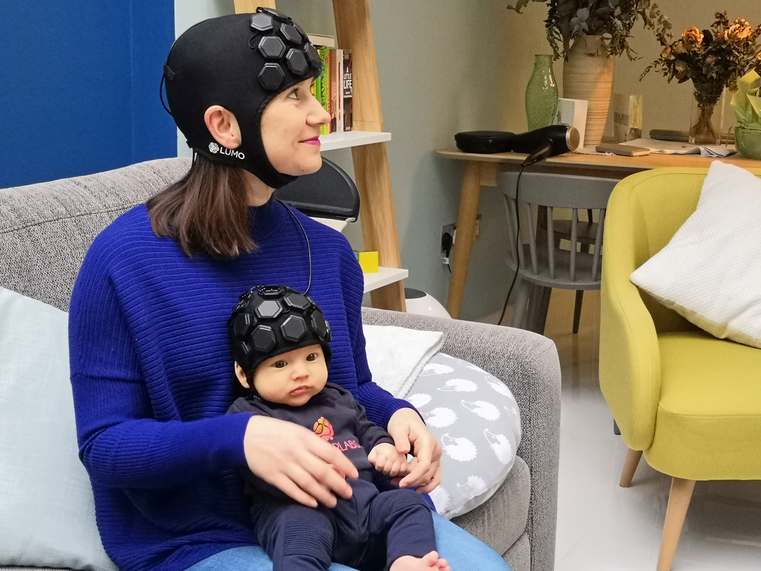
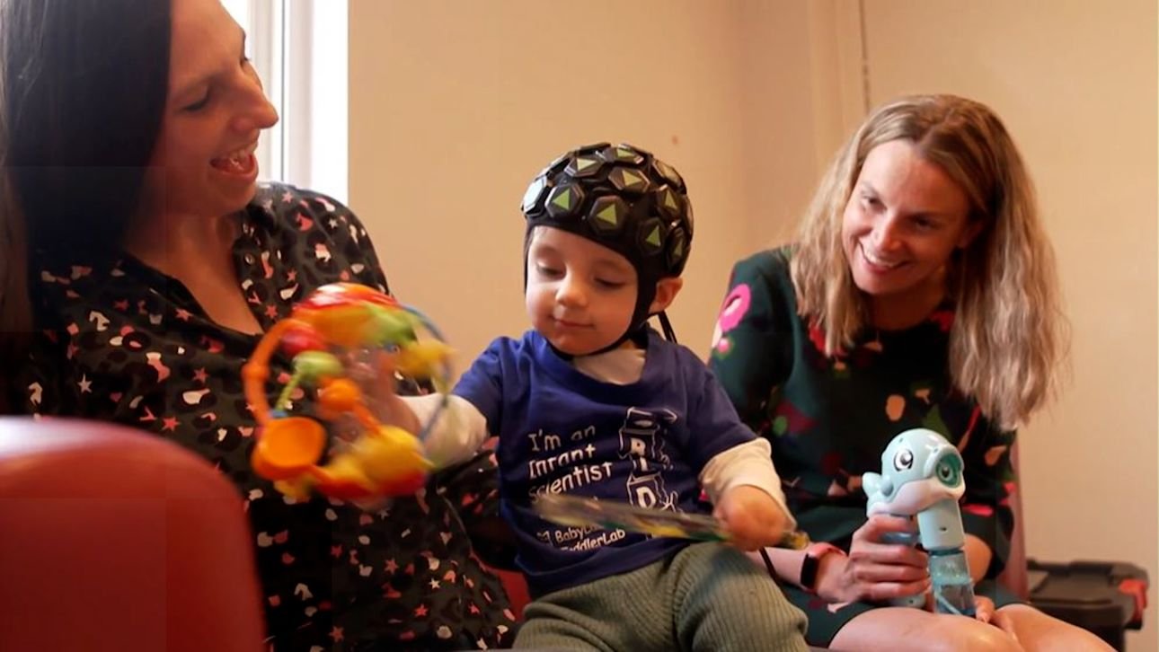
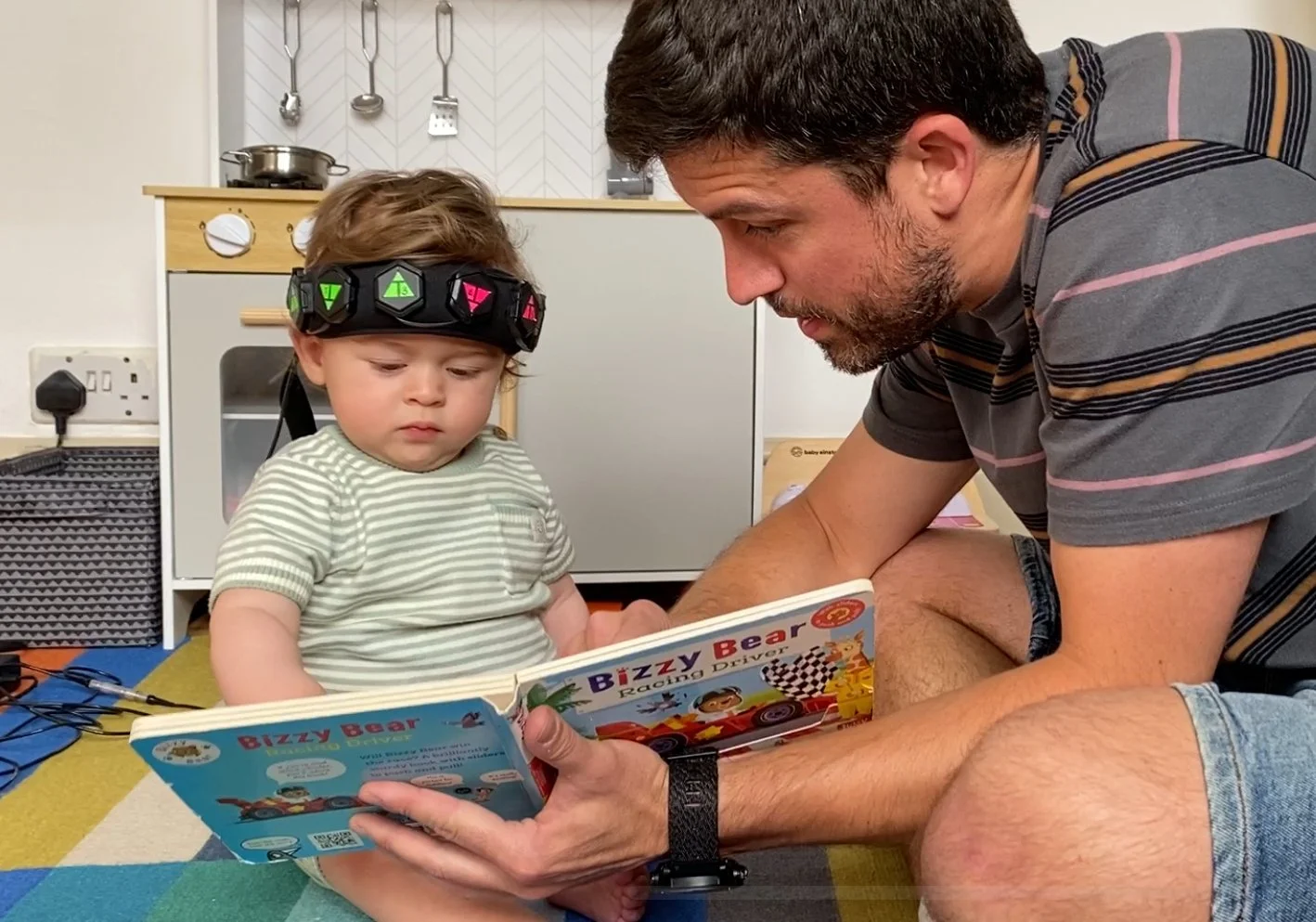
Child & Infant Studies
-
![]()
Whole-head Adult Imaging
-
![]()
Whole-head Infant Imaging
-
![]()
Clinical Settings
-
![]()
Complex Tasks & Environments
-
![]()
Neonatal Research
-
![]()
Real-World Environments
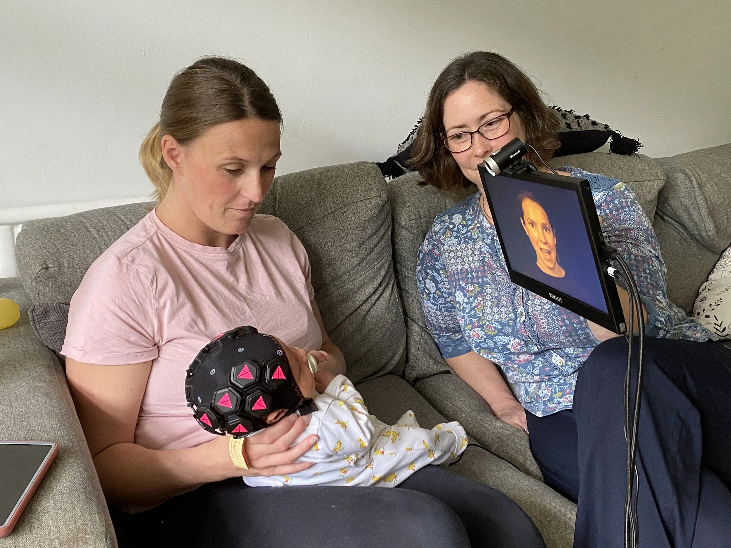


Real-world settings
LUMO is ultra compact and lightweight, making it perfect for naturalistic research.
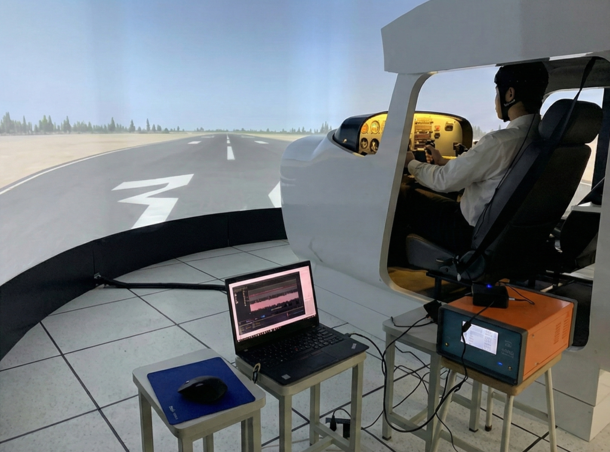
FIRST OF ITS KIND
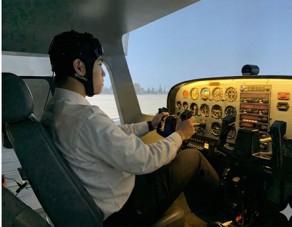
LUMO enables real-time detection of pilot fatigue - transforming aviation safety research using only 3 tiles
From the lab to the cockpit, LUMO’s compact wearable design makes high-quality brain monitoring possible even in a Cessna 172 flight simulator. Researchers at Nanjing University of Aeronautics and Astronautics used LUMO to monitor cerebral oxygenation during simulated flight tasks.
“Results show that the SDAE model established in our study has high identification accuracy, which can accurately identify different fatigue states of pilots. Identification of pilots’ fatigue status based on fNIRS has important practical significance for reducing flight accidents caused by pilot fatigue.” - Pan et al (2022)
Expanding the reach of neuroscience - the first demonstration of wearable HD-DOT in a non-laboratory environment
Researchers from University College London and the University of Cambridge used a 24 tile LUMO to investigate resting state functional connectivity networks in a home setting, during COVID-19 lockdown.
“This study represents the first demonstration on the use of wearable HD-DOT as a complimentary tool to fMRI for brain imaging in a non-laboratory environment.” - Uchitel et al (2022)
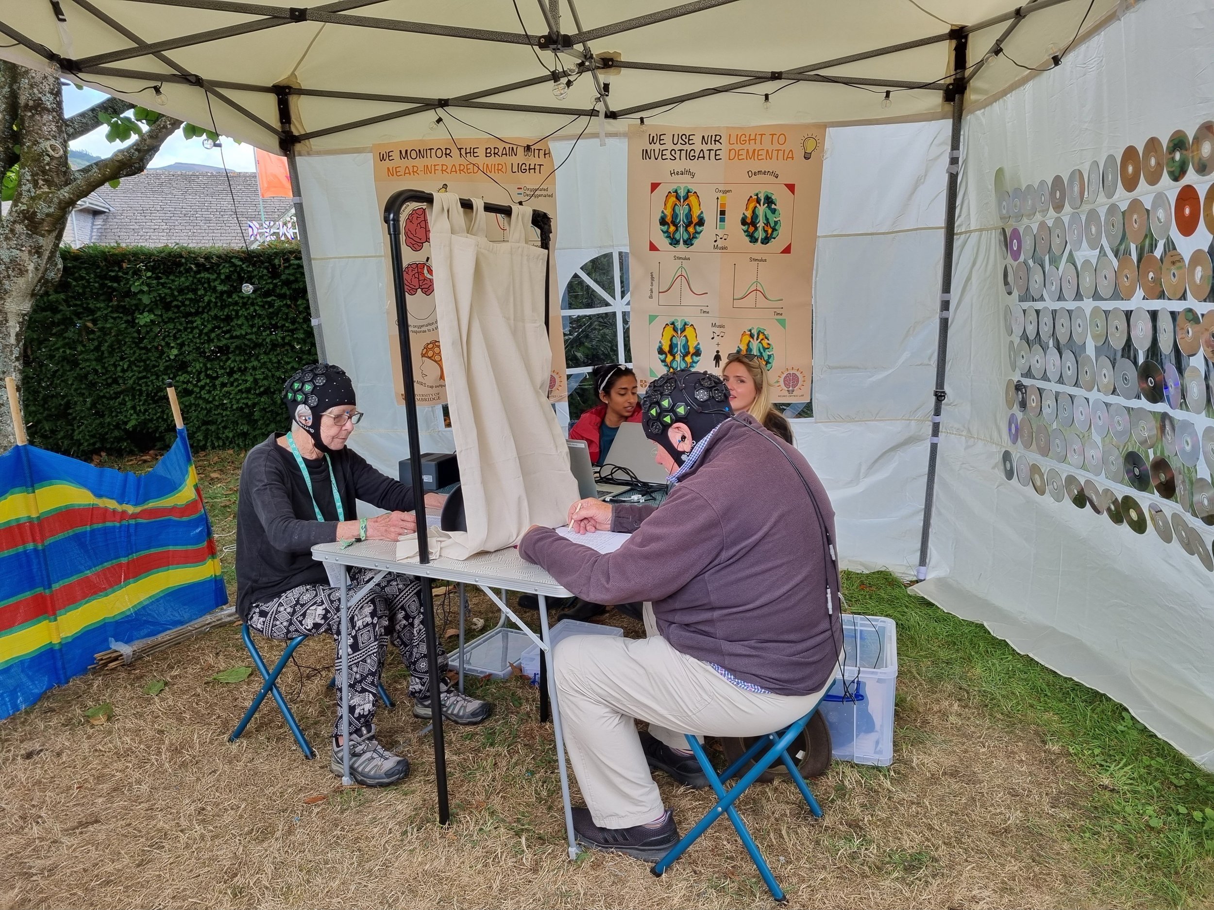
Clinical settings
With no bulky cables or fibres getting in the way and exceptionally fast setup time, LUMO is well suited to clinical environments and applications.
FIRST OF ITS KIND
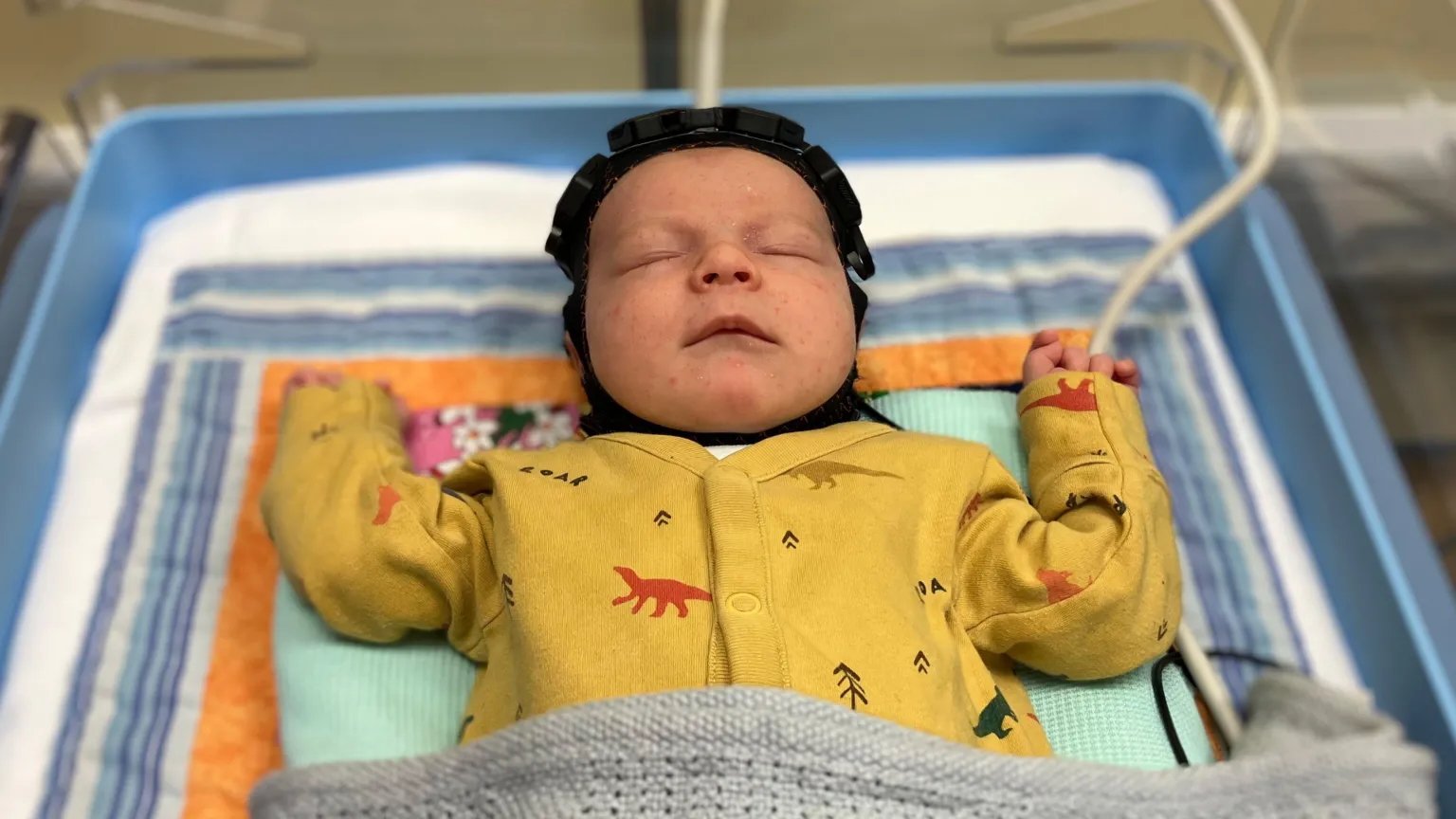
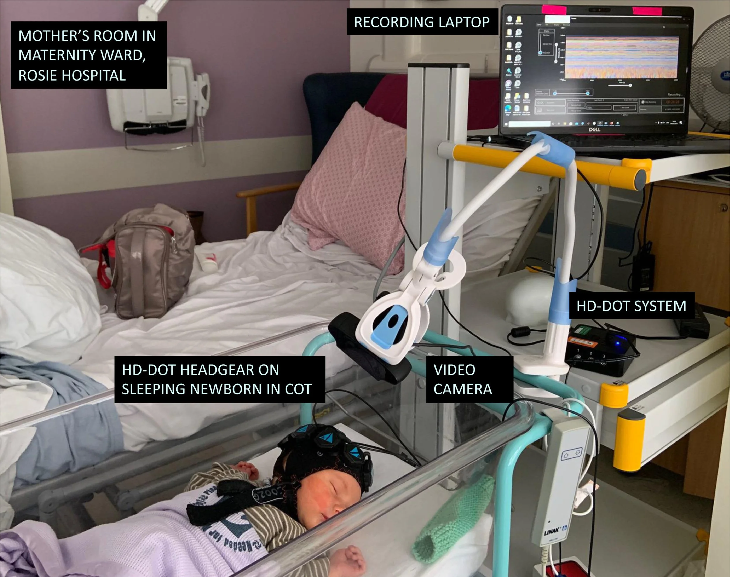
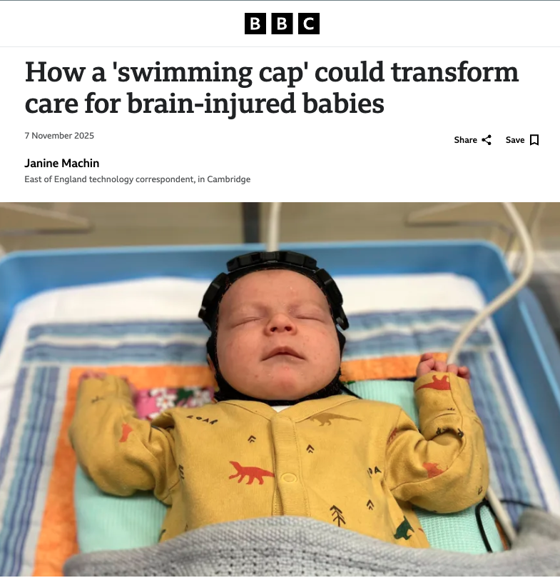
Bringing high-density brain imaging to the cot-side - neonatal neuroscience in a real clinical environment using 12 tiles
Researchers from University College London and the University of Cambridge used LUMO to study functional connectivity mapping in newborns during natural sleep, without the need for sedation or transport that other imaging methods require.
“[HD-DOT technology] overcomes many of the limitations of traditional, fibre-based and low-density fNIRS measurements. Driven by the development of this new technology, we have undertaken the first cot-side study of newborn infants using wearable HD-DOT in a clinical setting.” - Uchitel et al (2023)
FIRST OF ITS KIND
FIRST OF ITS KIND
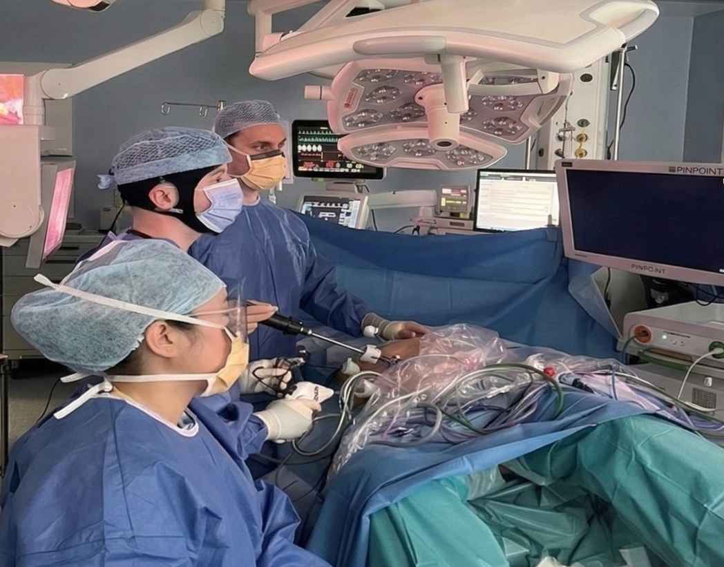
Using just 6 tiles to monitor cognitive workload in surgeons - enabling the first-ever fNIRS study during live surgery
In a groundbreaking application, researchers from UCD Centre for Precision Surgery, Mater Misericordiae University Hospital, and the University of Birmingham used LUMO to monitor cognitive workload during surgical procedures.
“fNIRS has previously been used to evaluate surgeons’ cognitive workload… in simulated scenarios but not previously in real-world surgical cases.” - McEntee et al (2025)
The first combined fUS-DOT neonatal imaging - a springboard for assessing functional connectivity at the whole-brain level
Researchers from Hospital Robert Debré, INSERM and the University of Cambridge, successfully combined a 12-tile LUMO HD-DOT system with functional ultrasound to capture whole-brain connectivity in neonates at the cot-side. This pioneering study demonstrated that both modalities can operate simultaneously without interference, laying the foundation for the comprehensive, early detection of neurodevelopmental disorders.
“fUS and DOT offer complementary brain coverage enabling comprehensive cot-side brain connectivity analysis.” - Faure et al (2025)
FIRST OF ITS KIND
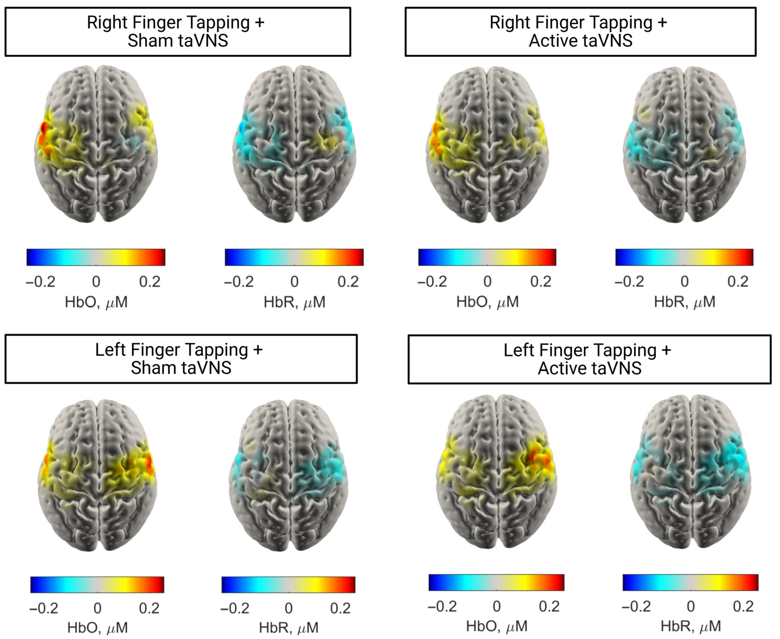
Real-time cortical monitoring during concurrent vagus nerve stimulation - demonstrating the feasibility of HD-DOT as a biomarker in stroke rehabilitation
Researchers from the University of Sheffield successfully recorded cortical haemodynamic responses across the sensorimotor cortex during concurrent motor tasks and non-invasive vagus nerve stimulation. LUMO can reliably detect localised task-related activation, showing that targeted HD-DOT could unlock a new way to measure the real-time impact of stimulation on stroke recovery.
“This study provides the first evidence of the feasibility of taVNS with HD-DOT and the first use of functional neuroimaging with synchronous taVNS and motor tasks.” - Baig et al (2026)
Whole-head high-density imaging
LUMO is the world’s lightest, fastest whole-head HD-DOT system.
Explore whole-head applications below.
FIRST OF ITS KIND

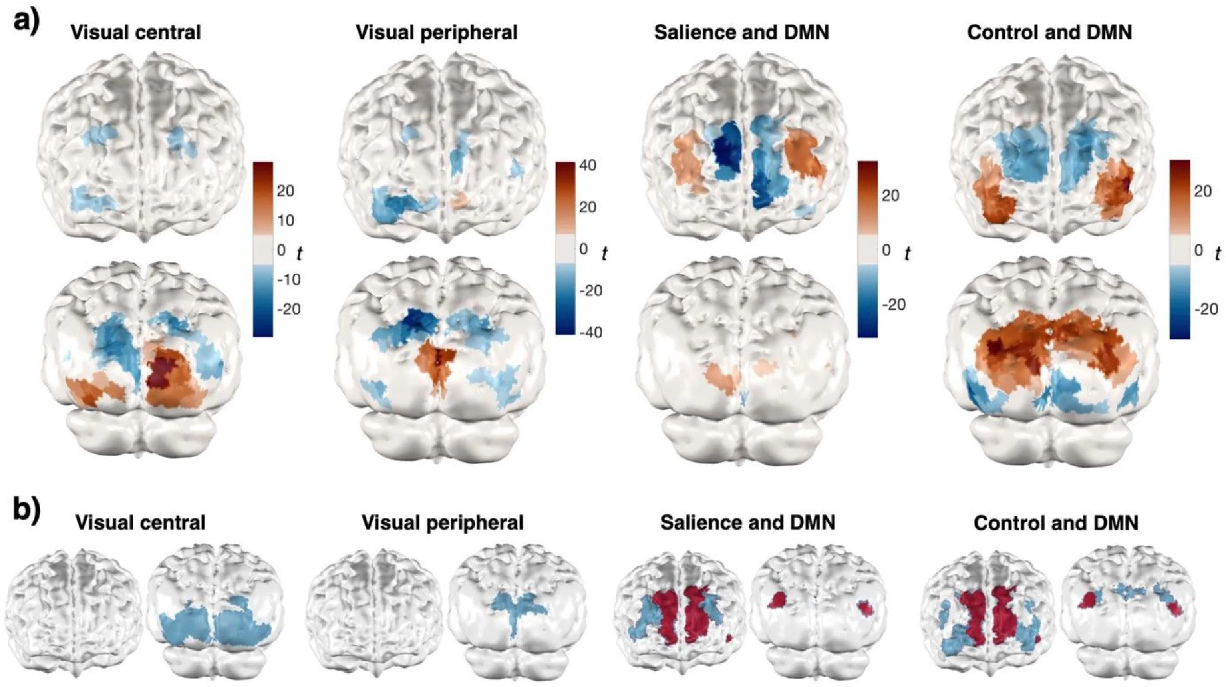
Using a 33 tile LUMO system to conduct the first-ever whole-head optical imaging of the awake infant brain
Researchers from Birkbeck, University of London utilised a 33-tile, whole-head LUMO array to measure cortex-wide brain activity in 16 awake infants (5–7 months old) during a dynamic audio-visual social processing task. By successfully capturing high-density data across the entire scalp, the team mapped functional responses in regions far beyond the limited field-of-view of previous infant neuroimaging tools.
“Combined with its portability, ease of set-up, and high tolerance among infants, whole-head HD-DOT offers the ability to obtain cortex-wide measures across an unprecedentedly large field-of-view outside of a conventional scanner setting.” - Collins-Jones et al (2024)
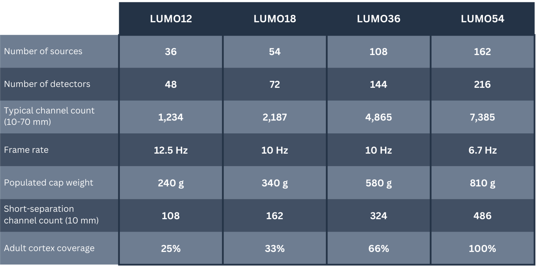
Example configurations
Every cap is custom-designed and built to order based on your specific regions of interest. Dock arrays can be partially or fully populated with tiles, enabling single or multi-region imaging. Compare key specifications across different system configurations in this table.
Contact us for a tailored quote or system demonstration.