
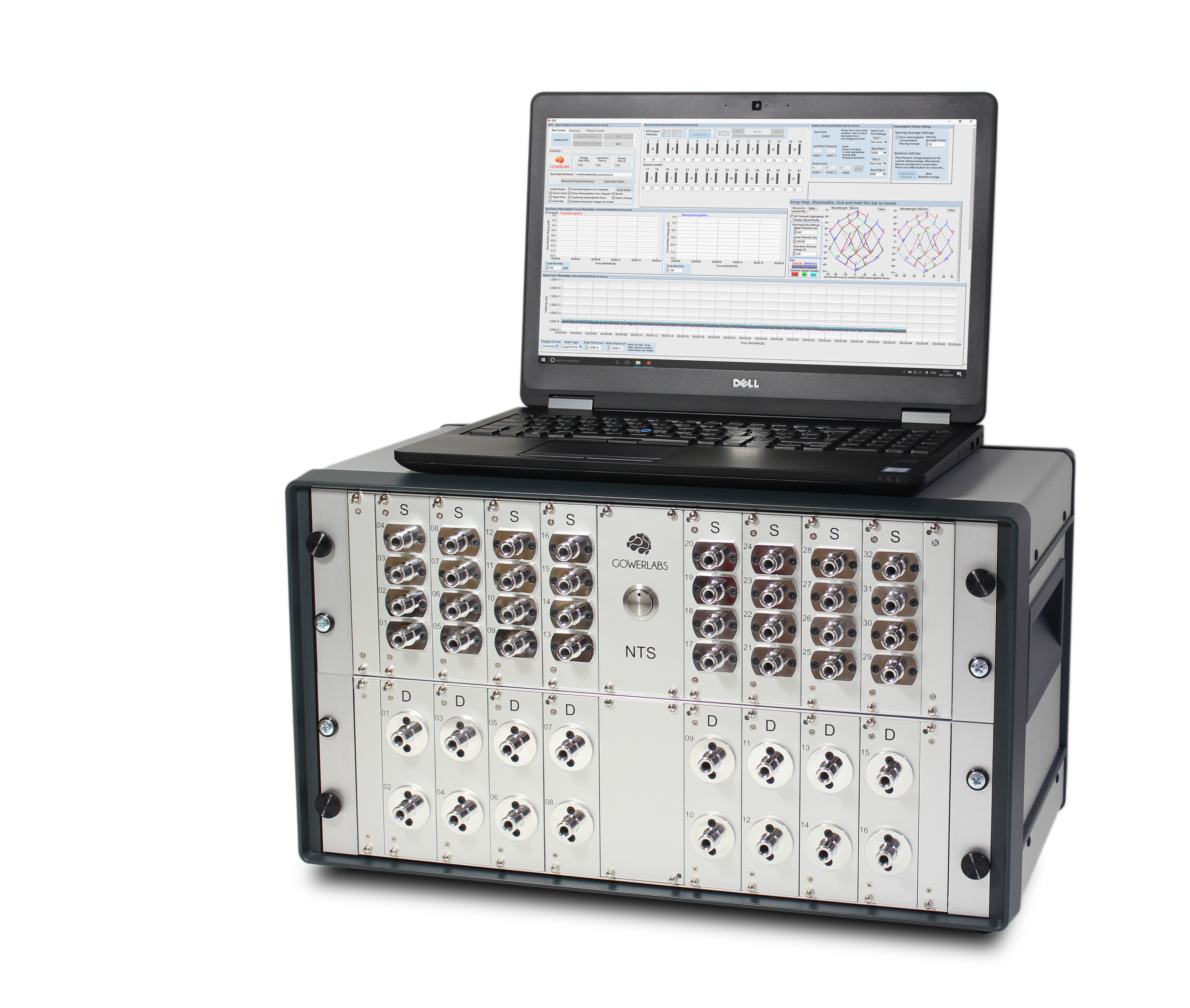
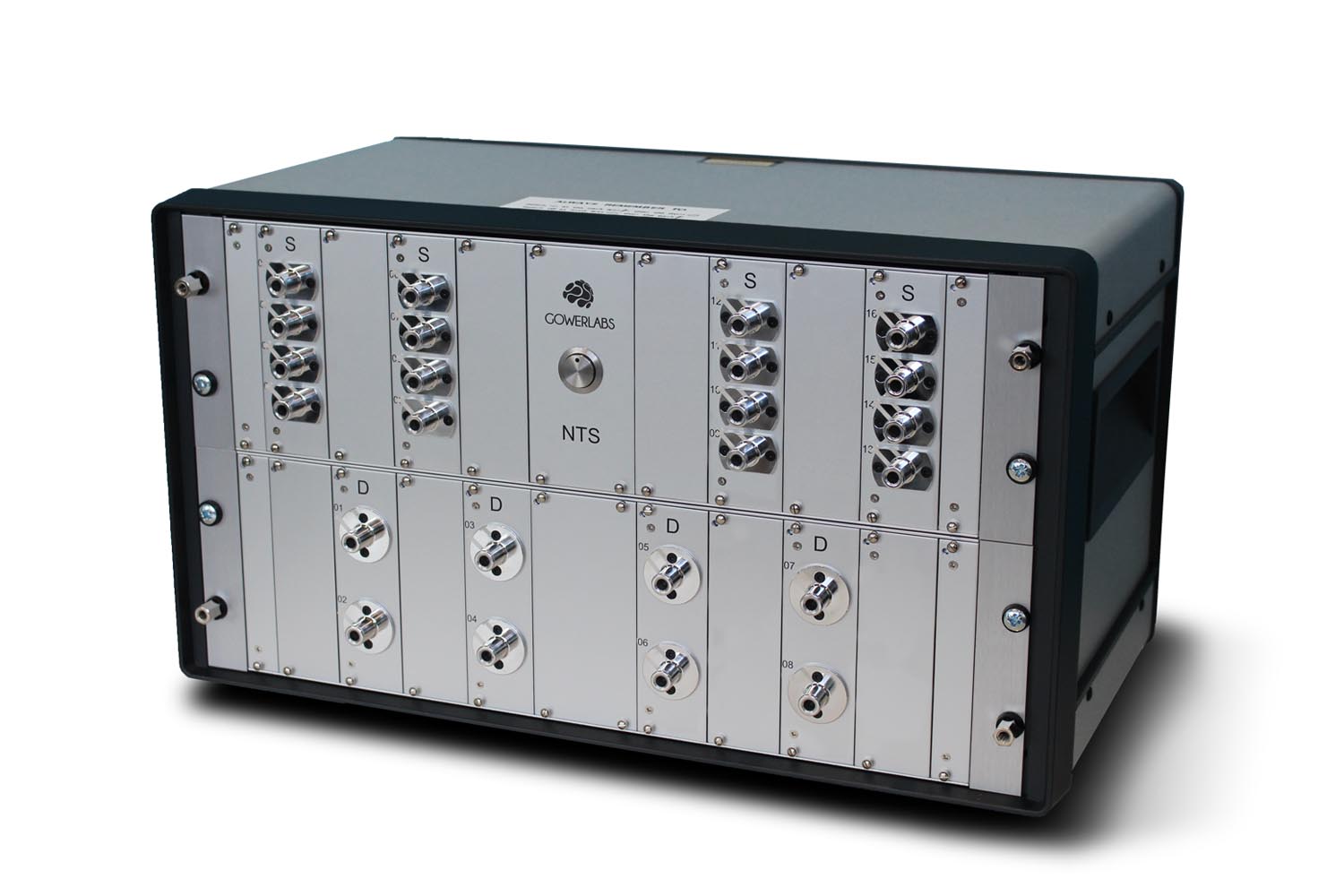
The NTS fNIRS System
Launched commercially in 2013, the NTS was designed to be flexible, smaller and less complex than existing optical topography systems to enable application to a wide variety of experimental and clinical investigations.
This advanced brain-mapping system, designed and built by Europe’s leading biomedical optics research group, performs non-invasive imaging of brain function in real time.
-
The NTS fNIRS system uses light to image changes in blood volume and oxygenation. This system was developed from 25 years of research excellence at UCL's Biomedical Optics Research Laboratory (BORL).
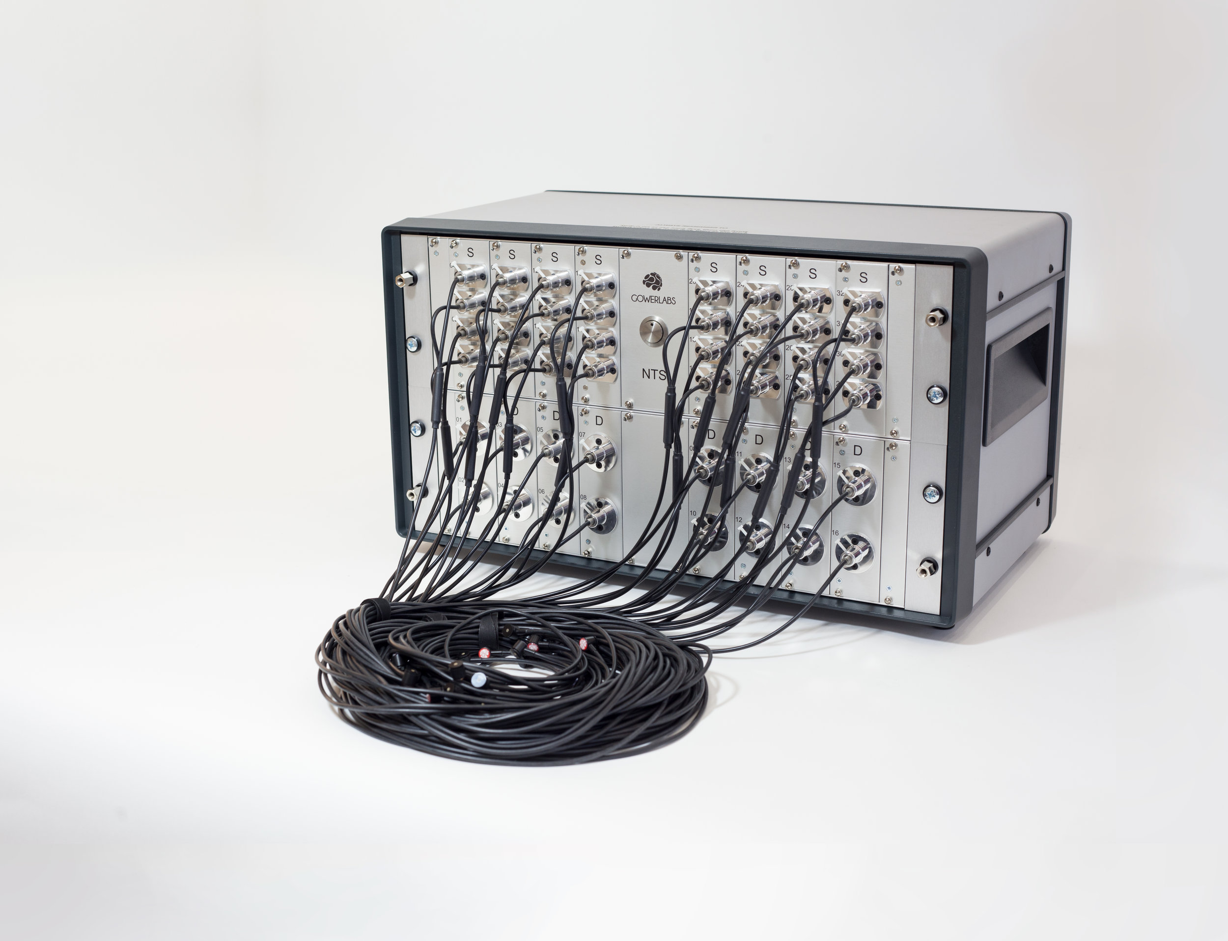
Two wavelengths of infrared light are injected at different points on a subject’s head, and the diffused light is measured at separate detection points. The system allows flexible configurations to study adults, children or neonates, thanks to an illumination technique that allows completely adaptable positioning of light sources and detectors. A variety of headgear options enable users to secure fibres comfortably to a subject’s head, and the entire system is controlled via an easy-to-operate graphical user interface.
Our article in the Review of Scientific Instruments describes the original NTS fNIRS System in more detail.
Click here for more information about Diffuse Optical Tomography, a key technology behind the NTS fNIRS system. For more information about and images of the system in use, see case studies.
-
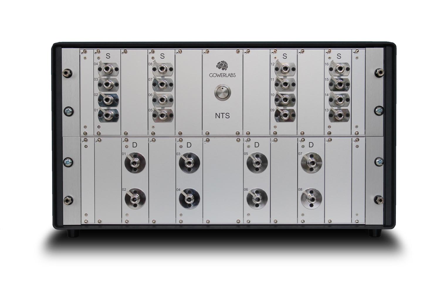
Laser diodes provide source light at 780 nm and 850 nm. Other wavelengths are also available
Modular expandable design
Our versatile and easy-to-use software package allows for a wide range of NIRS and fNIRS experimental applications
Any combination of sources and detectors are available. Common configurations include:
mini system: 6 sources, 4 detectors
half system: 8 sources, 8 detectors
full system: 16 sources, 16 detectors
-
Completely non-invasive
Ideal for infants, children and vulnerable subjects
No ionising radiation
Portable
Easy-to-use
Very low running costs
High spatial and temporal resolution
CE marked
To read about the NTS Headgear and Fibres, or to see some of the brilliant applications of the system, click the links below.

NTS Headgear
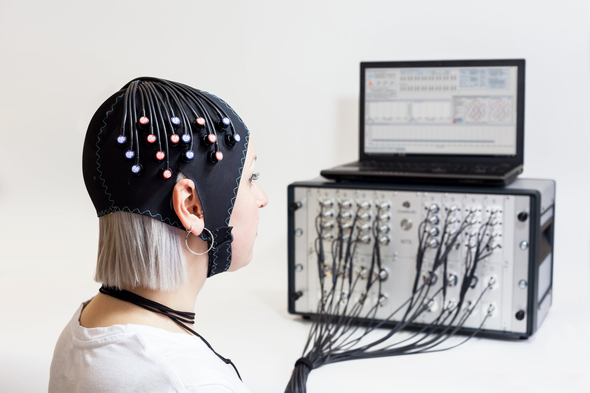
Gowerlabs' extensive research experience has resulted in a range of fNIRS headgear options. We have worked closely with our users to design and develop bespoke headgear solutions that meet the specific demands of their research programmes.
Headgear
fNIRS headgear must be comfortable, reliable, and adaptable. At Gowerlabs, our headgear solutions come in two principle styles: our specially designed EasyCaps, and CBCD’s bespoke headgear designed specifically for infant subjects.
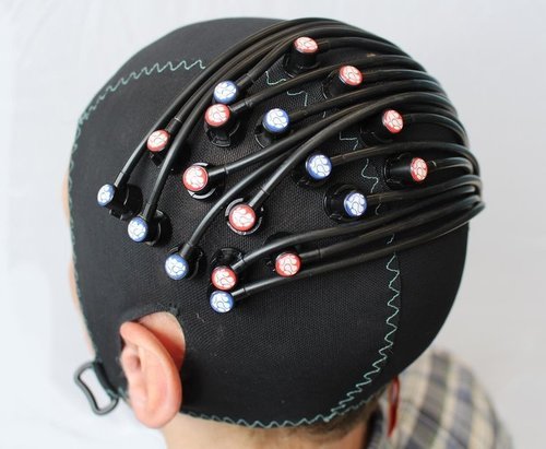

Our standard headgear is based on EasyCaps caps (EasyCap GmbH, Germany): a flexible, anatomically registered head-cap system originally developed for EEG research. EASYCAP caps are available in a wide range of sizes and can be used with all ages, from newborn infants to adults. The caps are comfortable, washable, and easy-to-apply with the Gowerlabs NTS fNIRS system. Because they are designed around the EEG 10-20 coordinate system, these caps also facilitate the direct spatial registration of fNIRS measurements with the cortical anatomy. More information about EASYCAP caps can be found here.
The second format is available via our partners at the Centre for Brain and Cognitive Development (CBCD, also known as Babylab) at Birkbeck, University of London. Babylab designs and manufactures fNIRS headgear for infants and toddlers based on their extensive experience applying Gowerlabs' fNIRS technology to study cognitive development. More information about the CBCD fNIRS headgear can be found here.
Short Separation Channels
In recent years, research has shown that short-separation fNIRS channels can be used to significantly improve the accuracy and reliability of fNIRS measurements.
The flexibility of the Gowerlabs NTS fNIRS system allows users to incorporate multiple short-separations measurements into their array design, resulting in higher-quality fNIRS data and more accurate interpretation.
Fibres
The NTS employs high-quality, lightweight, and flexible glass fibre bundles that provide excellent optical coupling and transmission. Our fibres are constructed from thin-cladded optical fibre to provide a maximum packing fraction, and thus increase light collection.
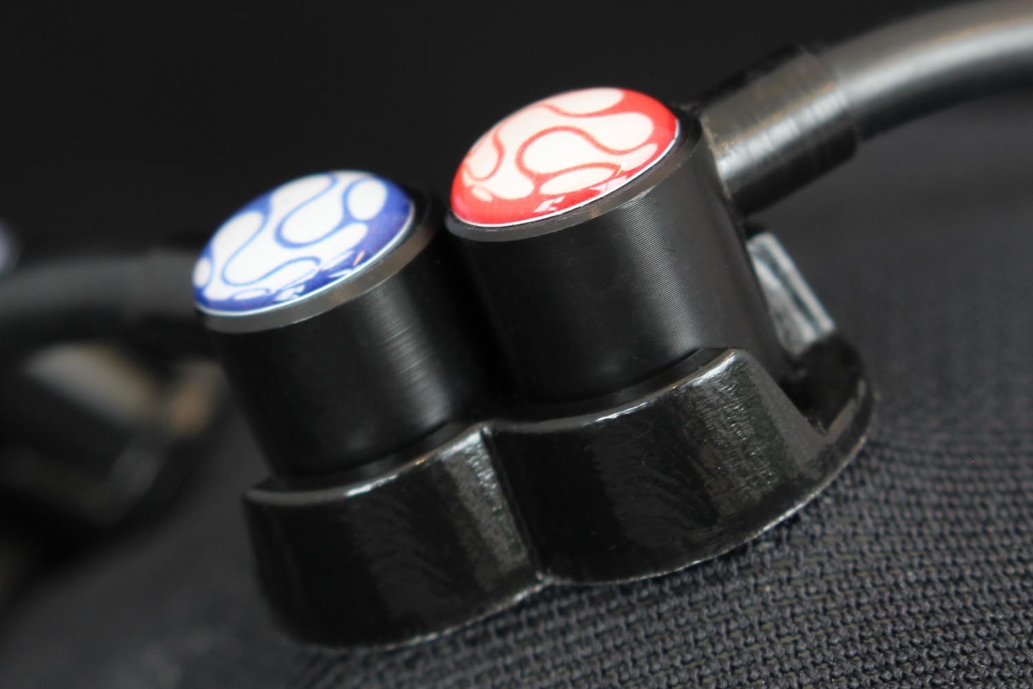
Source fibre bundles are bifurcated and incoherent to allow for the transmission of two wavelengths of light to the exact same point on the scalp. Gowerlabs’ fibre bundles come in three different designs.

Infant Fibre Bundles
Each infant optical fibre bundle has a flat, wipeable surface that is designed to minimise the pressure applied during fNIRS recording. These fibres work with our infant foam couplers, which provide a padded, opaque coupling surface that helps to maximise subject comfort.

Adult Fibre Bundles
Gowerlabs adult fibres bundles include extra features to improve fNIRS signal quality in the presence of thicker hair. Each bundle is coupled into a specialised adapter that fixes into the EasyCap in conjunction with a foam coupler that ensures fibre stability and subject comfort.

Adult+ Fibre Bundles
Our adult+ fibre bundles have an extended fibre tip and are designed to work with Gowerlabs’ spring-loaded fibre adapters. This has shown a greater than 10x improvement in signal-to-noise ratio in subjects with thick, dark hair while still remaining comfortable.
NTS Case Studies
We are pleased to feature here a selection of the exceptional research being undertaken by NTS users.
Centre for Advanced Reconstruction of Extremities (CARE)
At the Centre for Advanced Reconstruction of Extremities (CARE) at Sahlgrenska University Hospital (Gothenburg, Sweden), researchers are using the NTS fNIRS system and fMRI to map patterns of brain activity after reconstructive upper limb surgery in individuals with tetraplegia.
For more information about CARE and their research, please contact Dr. Lina Bunketorp Käll (lina.bunketorp@neuro.gu.se).

In upper limb tetraplegia reconstructive surgery, a functionally intact muscle is detached, rerouted, and reattached to a non-working muscle to replace its original function. By restoring this aspect of hand function, patients regain the ability to grip objects again hugely increasing their quality of life. Through recording the haemodynamic response to tasks such as key-pinching and elbow flexing, CARE is studying the resultant cortical plasticity in the brain as movement is restored.
After extensive discussions between Gowerlabs and CARE, a DOT array was designed that could image the primary motor cortex/primary somatosensory cortex (M1/S1), supplementary motor area (SMA), and premotor cortex (PMC). The array consists of 16 dual-wavelength sources and 16 detectors, including two short separation channels to remove scalp contamination in the signal.
During the experiment, both the control group and the patient group were asked to use their right hand to perform isometric key pinch and elbow flexor tasks. Each task was repeated five times, interspersed by periods of rest. The data from the NTS was analysed and reconstructed into images with the help of researchers at UCL.
Preliminary DOT results suggest that the control group demonstrates a clear lateralisation of the movement tasks to the contralateral hemisphere. In contrast, for the tetraplegic group the haemodynamic response to both tasks was highly bilateral. The findings suggest that upper limb tetraplegia reconstructive surgery induces cortical plasticity of the M1/S1 region to restore thumb function and enable gripping.


NeoLAB
Founded in 2013, neoLAB is a collaborative research partnership between UCL and Cambridge University. Based in the Evelyn Perinatal Imaging Centre (EPIC) at the Rosie Hospital, Cambridge, the group is comprised of physicists, computer scientists, and engineers from UCL collaborating with clinicians and researchers from Cambridge.
Located directly below the Neonatal Intensive Care Unit (NICU) at the Rosie Hospital, the EPIC was purpose-built to provide researchers unparalleled access to a wide range of imaging devices for investigating neonatal brain function, including electroencephalography (EEG), fNIRS, and magnetic resonance imaging (MRI). This makes it one of the best locations in the world to study baby brain development.
For more information about neoLAB’s research and the facilities at the EPIC, please visit their website.
The NTS fNIRS system is used by doctors and other researchers to investigate brain function in both healthy and sick infants. Because of the portability of the system, it can be wheeled right up to the cot of a baby in the NICU with ease. This way the patient is not disturbed and their clinical care can continue without interruption.
NeoLAB researchers have used the system to successfully scan the brain activity of sick infants when they are experiencing seizures and/or seizure-like events (such as burst suppression). This was made possible by integrating optical imaging with EEG. The following video shows the hemodynamic response to multiple seizures in an infant brain over the course of 5 minutes (DOT images are on top, EEG signals on bottom). Globally, the brain primarily shows a short increase in total haemoglobin concentration before experiencing a profound and long-lasting decrease. This corresponds to a significant reduction in blood volume where the brain is below normal oxygen levels for several minutes (see Video 1 below).
Not all infants with brain damage display such obvious seizures on EEG. Others may exhibit only subtle changes in their EEG (such as burst-suppression) which can be difficult to detect, but this information can be complemented with DOT imaging. Several infants experiencing burst-suppression events were imaged by neoLAB, and upon compiling the data it was clear that there were dramatic swings in total blood volume within the brain in response to these brief periods of abnormal activity (see Video 2 below).
Video 1
Video 2
More details about neoLAB’s work imaging seizures in the infant brain can be found in the following publications:
M. Chalia, C. Lee, L.A. Dempsey, A.D. Edwards, H. Singh, A.W. Michell, N.L. Everdell, R.W. Hill, J.C. Hebden, T. Austin, R.J. Cooper. “Hemodynamic response to burst-suppressed and discontinuous electroencephalography activity in infants with hypoxic ischemic encephalopathy.” Neurophotonics 3(3), 031408, 2016.
H. Singh, R.J. Cooper, C. Lee, L.A. Dempsey, A. Edwards, S. Brigadoi, D. Airantzis, N.Everdell, A. Michell, D. Holder, J.C. Hebden, T. Austin. “Mapping cortical haemodynamics during neonatal seizures using diffuse optical tomography: A case study.” NeuroImage: Clinical 5, 2014.
R.J.Cooper, J.C.Hebden, H.O’Reilly, S.Mitra, A.Michell, N.L.Everdell, A.P.Gibson, T. Austin. “Transient haemodynamic events in neurologically compromised infants: A simultaneous EEG and diffuse optical imaging study.” Neuroimage 55:1610-6, 2011.
LonDownS Consortium
At UCL, the Division of Psychiatry and the Department of Medical Physics and Biomedical Engineering are collaborating to study functional changes in the brains of people with Down Syndrome as they age.
For more information please see the website of the LonDownS Consortium or contact Rosalyn Hithersay at r.hithersay@ucl.ac.uk
Whilst the life expectancy of those with the condition has risen dramatically over the past 50 years, it is now apparent that they suffer disproportionately from Alzheimer’s disease. To address the lack of functional neuroimaging research in the Down Syndrome population, UCL is using fNIRS and DOT to study executive function in people with Down Syndrome of all ages. Decline in executive function is a possible early feature of Alzheimer’s in Down Syndrome and the research team hopes to monitor executive function abilities to better understand the corresponding haemodynamics in the brain.
The frontal cortex is highly involved in executive function, showing activation during tasks requiring rule learning, shifting, and inhibition. With the help of Gowerlabs, UCL researchers developed a DOT array covering the frontal cortex and superior temporal cortex with 16 sources and 16 detectors, for a total of 44 channels. Data was acquired with the NTS fNIRS system during several different paradigms designed to activate the frontal cortex, including a go/no-go task, picture Stroop task, verbal fluency task, and dimensional change card sort task. To date, pilot studies with 9 adults with Down Syndrome and 12 control participants have been completed and a larger study is now underway.
To our knowledge, this is the first study to use fNIRS with adults with Down Syndrome. Crucially, this research is possible because fNIRS allows more natural movement than other imaging modalities such as fMRI. The subjects tolerated the headgear and Gowerlabs fibre optics well, making it possible to study executive function in this population. Gowerlabs is excited to see where this new study leads and will continue to provide support for the research as it progresses.


UCL Institute of Cognitive Neuroscience
Researchers at the UCL Institute of Cognitive Neuroscience and Babylab (Birkbeck College, London) are collaborating to study the impact of early speech and language experience on the neural representation of language in infancy.
For more information, please find Dr. Evelyne Mercure’s contact details on the UCL Institute of Cognitive Neuroscience website.

Led by PI Dr. Evelyne Mercure, the group is interested in comparing hearing infants of deaf mothers who use a sign language as their dominant language (HoD infants) with hearing infants of hearing mothers (HoH infants). This is of interest because despite having normal hearing, HoD infants have a different early experience of speech and language to that of HoH infants. Firstly, since deaf mothers are more likely to use signing than auditory speech, their infants are likely to have reduced exposure to auditory spoken language. Secondly, the experience of HoD infants includes both a visual language (e.g. British Sign Language (BSL)) and an auditory language (e.g. English). The team decided to compare HoD infants to bilingual HoH individuals since this latter group is also exposed to two languages (both auditory). They also included a control group comprised of monolingual HoH infants. They hypothesised that the experience of HoD infants will influence their neural representation of language.
All subjects had fNIRS data acquired using the Gowerlabs NTS system. Headgear containing 38 source-detector channels was developed at Babylab and designed to image the temporal and temporoparietal areas of the brain. The latter area was included because this part of the brain has previously been seen to play an important role in processing sign language. There were 38 channels in the array with 2 cm source-detector separations. Dr. Mercure commented that she is very happy with fNIRS as a tool to scan baby brain function in comparison to her previous experiences with EEG and fMRI. Particularly noteworthy is the fact that she can scan the infants when they are awake and behaving naturally. She also attributes her higher success rate to her subjects’ increased tolerance to Gowerlabs specially designed headgear.
Each of the three subject groups were shown videos of people telling short stories in four different languages, and their functional responses were then compared. The languages were: English (familiar spoken language), French (unfamiliar spoken language), British Sign Language (familiar sign language to HoD infants), and French Sign Language (unfamiliar sign language). The aim was to assess how the brain represents spoken and signed languages, as well as familiar and unfamiliar languages. To date, the team has scanned around 100 infants with the NTS fNIRS system for this study!
Preliminary results from the temporal channels suggest that spoken language causes a strong activation in the temporal cortex across all subjects. For the HoH monolinguals, the response is highly bilateral, but in the HoD infants it is more restricted and left-lateralised; whilst in the HoH bilinguals it is more right-lateralised. The rich data-set collected by this group is still being analysed, and we can expect many more interesting results from this study in the near future.

Global fNIRS
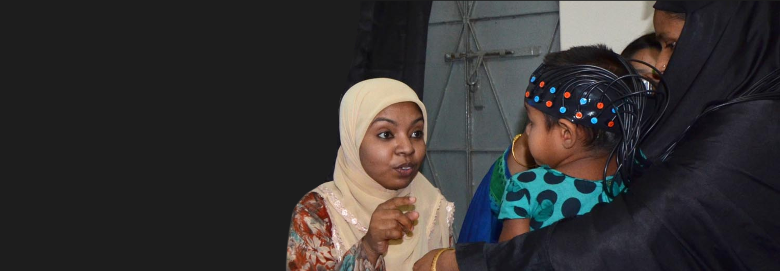
BRIGHT is part of a larger initiative known as Global fNIRS, a multidisciplinary project dedicated to advancing global health initiatives through the use of fNIRS. Their goal is to provide tools which supplement current methods for assessing cognitive function of the developing brain in resource-poor settings. With this goal in mind, a related project led by Professor Charles Nelson (Boston Children’s Hospital) is currently in progress in Dhaka, Bangladesh, where our imaging system is being used to record data from children, to predict future cognitive function. Again, the portability and ease-of-use of the NTS fNIRS system made it possible to successfully run these experiments in a country with limited resources. Gowerlabs looks forward to seeing where Global fNIRS will go next!
The BRIGHT Project
The Brain Imaging for Global Health (BRIGHT) Project is a collaborative research effort led by a team of interdisciplinary researchers from UCL, Birkbeck (University of London), the Medical Research Council (MRC) Units in the Gambia and Cambridge, King’s College London and Cambridge University Hospitals.
The project was funded by prestigious Phase I and Phase II grants from the Bill and Melinda Gates Foundation. BRIGHT project Director Clare Elwell gave a spotlight talk on Brain Imaging in Global Health at the Grand Challenges - Gates Foundation Annual Meeting 2016 (see Video 3).
Children raised in low-resource countries can be exposed to under-nutrition, social and environmental difficulties, increased risk of disease, as well as other issues relevant to cognitive development. The BRIGHT project is a longitudinal study following 200 Gambian and 60 UK infants from birth to 24 months of age. The aim of the project is to establish brain function-for-age curves of infants in both these cohorts. These and other data will provide an insight into the effects that living in a low-resource context can have on infant development.
While there is a large amount of research that highlights the detrimental impact that undernourishment and poverty have on infant development, not much is known about the neural basis of this. The BRIGHT Project intends to shed some light on this research area by not only tracking development through behavioural assessments, anthropometric measures, family, and environmental questionnaires, but also through the implementation of neuroimaging techniques. A primary aim of the BRIGHT project is to collect longitudinal functional brain response data using fNIRS (using the NTS fNIRS system) and EEG. The BRIGHT Project conducted the first ever functional brain imaging of infants in Africa!
The simplicity, portability, and ease-of-use of the NTS fNIRS system were crucial to the success of the Phase I pilot study for the work in Keneba. A significant factor contributing to this success stems from extensive work undertaken over the last ten years by Sarah Lloyd-Fox and Anna Blasi at the Centre for Brain and Cognitive Development at Birkbeck to develop a robust headgear solution suitable for infants (see fNIRS headgear and the NIRS CBCD Headgear Plugin webpage). The combination of this CBCD headgear and the NTS fNIRS system allowed the team to set up and complete a study within a few hours of arriving at the MRC Unit in The Gambia, on the very first trip there.
The CBCD headgear solution incorporates the Gowerlabs fibre optics, prevents slippage against the head (very useful when working with active children), is quick and easy to apply, and suits many different head shapes and sizes for the varied age cohort being studied.


Video 3
For more information about BRIGHT and Global fNIRS, please visit http://www.globalfnirs.org and follow twitter updates from the group @globalfnirs. Early results from the fNIRS imaging experiments in the Gambia can be found in the following publications:
S. Lloyd-Fox, K. Begus, D. Halliday, L. Pirazzoli, A. Blasi, M. Papademetriou, M.K. Darboe, A.M. Prentice, M.H. Johnson, S.E. Moore, C.E. Elwell. “Cortical specialisation to social stimuli from the first days to the second year of life: A rural Gambian cohort.” Developmental Cognitive Neuroscience, 2016. doi: 10.1016/j.dcn.2016.11.005.
S. Lloyd-Fox, M. Papademetriou, M.K. Darboe, N.L. Everdell, A.M. Prentice, S.E. Moore, C.E. Elwell. “Functional Near Infrared Spectroscopy (fNIRS) to assess cognitive function in infants in rural Africa.” Nature Scientific Reports (4:4740), 2014. doi: 10.1038/srep04740.


Birkbeck’s Centre for Brain Development and Cognition (CBCD)
The Centre for Brain and Cognitive Development (CBCD) was founded in 1998 at Birkbeck, University of London and is directed by Professor Denis Mareschal. It has grown steadily and is now internationally recognised as one of the leading centres of its kind in the world. The work of CBCD members is characterised by its use of converging methods (behavioural testing, eye tracking, ERP, EEG, NIRS, optical imaging, EMG, computer modelling, functional and structural MRI), and by its theory-driven programmes of empirical research on visual, cognitive, and language development in human infants, children, adolescents and adults.
For more information about this project please contact Carina de Klerk (c.deklerk@bbk.ac.uk), or visit the CBCD website.
Human adults have a tendency to spontaneously and unconsciously copy or ‘mimic’ others’ behaviours, including their postures, gestures, facial expressions, and emotions. This mimicry plays an important role in social interaction. For example, it has been shown to increase liking between strangers, and makes our interactions smoother and more enjoyable. However, little is known about when and how mimicry develops in humans. It is believed that some form of mimicry starts early in infancy, with new-borns mimicking their parents’ facial actions; however these findings are controversial. What is clear is that mimicry increases with age, and that by around 3 years of age, children seem to show the kind of spontaneous mimicry that we see in adults. To help address the knowledge gaps in this field, researchers at Birkbeck’s Centre for Brain Development and Cognition (CBCD) are investigating how mimicry develops from infancy to toddlerhood using electromyography (EMG) and the NTS fNIRS system.
The CBCD has been testing a cohort of sixty infants at six month intervals, from 4 months until 3 years of age, acquiring both EMG and fNIRS data. The EMG data gives an objective and sensitive measure of facial muscle movements during mimicry, whilst the fNIRS data can image brain activation. It has been suggested that the brain’s mirror neuron system plays a role in mimicry, and therefore, the team has focused on investigating brain regions that cover 1) areas that support mimicry (i.e. the superior temporal sulcus (STS) and inferior frontal gyrus (IFG)), and 2) areas that modulate mimicry (i.e. the medial prefrontal cortex (mPFC) and temporo-parietal junction (TPJ)). By using fNIRS to look at brain activation, the CBCD team can image awake infants and collect data that is both robust to movement and has better spatial resolution than electroencephalography (EEG) or magnetoencephalography (MEG).
Using an integrated EMG + fNIRS method, the CBCD will continue to assess mimicry responses and the underlying neural mechanisms in this cohort as these infants age. Their investigations will focus on how developmental factors such as sensorimotor experience, infants’ growing social cognitive abilities, and increasing brain connectivity influence the development and modulation of mimicry. Thus, we can expect more fNIRS results in the years to come with new and interesting cognitive paradigms.


