
Functional Near-Infrared Spectroscopy (fNIRS)
Functional Near-Infrared Spectroscopy (fNIRS) is a rapidly expanding neuroimaging technique. Gowerlabs systems are at the forefront of this growth, allowing human brain function to be studied in a non-invasive, easy-to-use, and portable way. Here, we describe the fundamental science behind our technology.

A Rush of Blood to the Head
Our brains are made up of billions of neurons and, like any cell, they require a ready supply of both oxygen and glucose in order to function. When neurons are active - firing signals in the forms of action potentials and passing information between one another through the release of neurotransmitter molecules - their metabolic demand increases. To ensure that they do not become deprived of oxygen or glucose, local blood vessels will quickly start to dilate in response to this increased activity. As a result, there is an influx of oxygenated blood to areas surrounding active neurons.
The relationship between neuronal activity and the localised response of the blood vessels is known as neurovascular coupling and, while its mechanisms are complex, the blood flow response to an increase in neuronal activity is reliable and well understood. Thanks to this, measurements of localised changes in cerebral blood flow provide an excellent proxy measurement of brain activity.
Seeing Red
Human tissues exhibit relatively low absorption of light in the red and near-infrared regions of the electromagnetic spectrum. As a result, near-infrared light can be transmitted through many centimetres of tissue. This is apparent in everyday life such as when you place your fingers over a white light they will appear to glow red (see Figure 1). The tissues in your hand absorb the violet, blue, green, yellow, and orange light, but leave much of the red light to pass through your fingers and reach your eyes.
Whilst red and near-infrared light are generally absorbed less than other colours, there are still many molecules in human tissues that do absorb light at these wavelengths. The principal absorber of near-infrared light in tissue is haemoglobin, the molecule which carries oxygen around the bloodstream. What's more, the oxygenated and deoxygenated forms of haemoglobin have distinctly different absorption spectra in the red and near-infrared range. To the naked eye, this has the effect of making blood that is loaded with oxygen appear a much brighter red than blood that has become deoxygenated.
The specific absorption spectra of oxygenated and deoxygenated haemoglobin is shown in Figure 2. The Gowerlabs NTS Optical Imaging System employs two wavelengths of near-infrared light (usually 780nm and 850nm though 685 nm and 850nm is also common) to allow changes in the concentration of both forms of haemoglobin to be measured. See section 'A Window to the Brain' for more details. Tissue spectra courtesy of UCL's Biomedical Optics Research Laboratory.

Figure 1
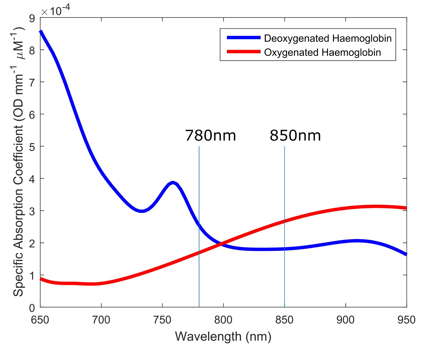
Figure 2
Figure 3 A typical fNIRS haemodynamic response function (HRF) measured by the Gowerlabs NTS Optical Imaging System. An increase in oxyhaemoglobin concentration (HbO) occurs in conjunction with a smaller decrease in deoxyhaemoglobin concentration (HbR), as oxygenated blood flows into the active brain region. HbT is the total haemoglobin concentration.
A Window to the Brain
These two critical properties, the relative transparency of human tissues to near-infrared light and the different absorption spectra of oxygenated and deoxygenated haemoglobin, provide us with an outstanding tool with which to investigate the human brain.
By transmitting near-infrared light at two wavelengths into the scalp and detecting the light that scatters back to the surface a few centimetres away, it is possible to measure the changes in concentration of oxygenated and deoxygenated haemoglobin in the brain. Due to the close relationship between neuronal activity and local blood flow described above, we can use near-infrared light to examine human brain function.
An increase in neuronal activity will result in a local increase in the volume of oxygenated blood. This corresponds to an increase in oxyhaemoglobin and, often, a simultaneous decrease in deoxyhaemoglobin. This is known as the Haemodynamic Response Function (or HRF).
Recommended Reading
Boas, David A., et al. "Twenty years of functional near-infrared spectroscopy: introduction for the special issue." NeuroImage 85 (2014): 1-5.
Scholkmann, Felix, et al. "A review on continuous wave functional near-infrared spectroscopy and imaging instrumentation and methodology." NeuroImage 85 (2014): 6-27.
Quaresima, Valentina, Silvia Bisconti, and Marco Ferrari. "A brief review on the use of functional near-infrared spectroscopy (fNIRS) for language imaging studies in human newborns and adults." Brain and Language 121.2 (2012): 79-89.
Lloyd-Fox, Sarah, Anna Blasi, and C. E. Elwell. "Illuminating the developing brain: the past, present and future of functional near infrared spectroscopy." Neuroscience & Biobehavioral Reviews 34.3 (2010): 269-284.